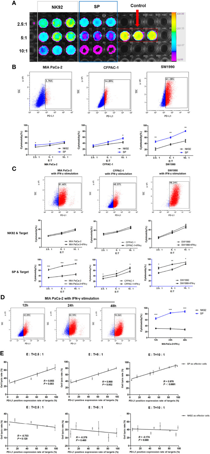
Figure 3.

Enhanced cytotoxicity of SP cells against both innate and IFN-γ-induced PD-L1-positive PDAC cells in a PD-L1 dose-dependent manner. (A) Cytotoxicity of SP or NK92 cells against SW1990 cells was measured by bioluminescence imaging after a 12-hour co-culture. (B) Expression of programmed cell death ligand 1(PD-L1) in three Pancreatic ductal adenocarcinoma (PDAC) cell lines (MIA PaCa-2, CFPAC-1, SW1990) was detected by FCM, and the lysis rate of SP and NK92 cells against each cell line was analyzed. (C) The three PDAC cell lines were stimulated with 5 ng/mL IFN-γ for 24 hours, the expression of PD-L1 in each cell line and the cytotoxicity of SP or NK92 cells against each cell line were detected. (D) MIA PaCa-2 cells were stimulated with 5 ng/mL IFN-γ for 12, 24, and 48 hours, to induce the expression of PD-L1 and the cytotoxicity achieved by SP or NK92 cells in co-cultures were assessed. (E) A correlation analysis between the cytotoxicity and target cell PD-L1 level. All summary data are indicated as mean ± SEM (n = 3). *P < 0.05, **P < 0.01 and ***P < 0.001.
