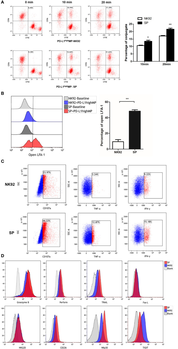
Figure 5.
FCM detection of SP cells conjugate formation, degranulation and cell death signaling transduction. (A) SP or NK92 cells were incubated with CFSE-labeled PD-L1highMP cells and assessed for the proportion of conjugate formation (CFSE and CD56 double positive counts). (B) SP or NK92 cells were assessed for the expression of LFA-1 in open-conformation following incubation with PD-L1highMP. (C) CD107a, TNF-α and IFN-γ expression of SP or NK92 cells were detected after a 6-hour incubation with PD-L1highMP. (D) SP or NK92 cells were assessed for expression of markers including granzyme B, perforin, TRAIL, Fas-L, NKG2D, CD226, NKp30, and TIGIT using FCM. For panels A and B, dummary data are shown as mean ± SEM (*P < 0.05, **P < 0.01, n = 3).

