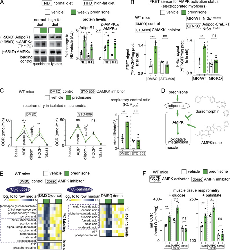
Figure 5.
Pulsatile prednisone activates the AMPK response to adiponectin in muscle. (A) Representative blots showing that intermittent prednisone increased the protein levels of total ADIPOR1 and phosphorylated AMPK in both normal and obese muscle. (B) FRET sensor in electroporated myofibers showed increased AMPK activity in muscle after a prednisone pulse. In WT mice, this effect was blunted by co-injection with the CAMKK inhibitor STO-609 (left). In line with GR-driven adiponectin elevation, this effect in muscle was blunted by fat-restricted GR ablation (right). (C) A prednisone pulse increased mitochondrial RCR in isolated muscle mitochondria and this effect was blunted by the CAMKK inhibitor STO-609. (D) Treatment effects on muscle AMPK were challenged with small-molecule AMPK modulators dorsomorphin (non-selective AMPK inhibitor) and AMPKinone (specific AMPK activator). (E) Dorsomorphin blocked the gain in nutrient oxidation and aerobic energy production induced by a prednisone pulse, shown here with 13C tracing in isolated muscle. (F) Muscle tissue respirometry showed that AMPKinone had additive effects on glucose- and palmitate-fueled respiration, while dorsomorphin abrogated prednisone-induced gains. Histograms, single values, and mean ± SEM. All panels report data verified in at least two independent experiments. n = 5 mice/group (A and F), n = 3 mice/group (B–E). *, P < 0.05; **, P < 0.01; ***, P < 0.001; one-way Welch’s ANOVA with Tukey multiple comparison; C (OCR curves): two-way ANOVA with Tukey multiple comparison for time points. Source data are available for this figure: SourceData F5.

