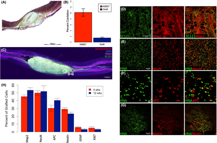
FIGURE 1.

Transplanted Human iPSC‐Derived sNPCs Fill the Lesion Cavity and Express Mature Neural Markers. (A) Human iPSC‐derived sNPCs were transplanted into T8 contusion SCI. Sagittal sections of Eosin Y/Nissl staining. (B) Cavitation analysis of the contusion site and transplanted cells in 1A. N2B27 is a media‐only control. (C) Immunolabelling of transplanted cells filling the lesion cavity. Rostral is left and caudal is right. Note: D‐G are representative examples. (D–G) Expression of neural markers Nestin, NeuN, Map2 and APC in transplanted human cells (HNA/SC121) twelve weeks post‐transplantation. (H) Quantification of expression of neural markers at 4 and 12 weeks post‐transplantation. Both Ki‐67 and GFAP expression are less than 5% after twelve weeks and approximately 90% of transplanted human cells are expressing mature neural markers. Data are represented as mean ± SEM
