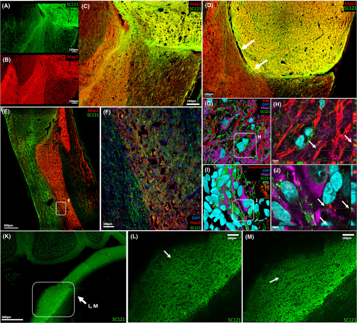
FIGURE 4.

Axonal Extension and Connectivity of iPSC‐derived sNPCs. (A‐C) SC121 and MAP2 staining identify mature transplant‐derived axons projecting into the host spinal cord. (D) SC121 and MAP2 staining indicate that successful axonal projection from the transplant into the host requires cell‐to‐cell contact. (E–F) SC121 and MAP2 staining also demonstrate that these axons extended in a rostral‐caudal direction in the rat spinal cord white matter, with axonal projections occasionally branching off and synapsing on host grey matter. (G‐H) SC121/MBP‐positive cells were located in close proximity and linearly aligned to host MAP2‐positive neurons, suggesting transplanted oligodendrocytes are myelinating host neurons. Arrows indicate 3D rendering of SC121/MBP‐positive cells in linear alignment with host MAP2‐positive cells. (I–J) Host MAP2‐positive neuron surrounded by SC121‐human synaptophysin‐positive puncta, suggests the establishment of synaptic contacts between the transplant and the host. Arrows indicate 3D rendering of a host MAP2‐positive neuron in close proximity to SC121‐human synaptophysin‐positive puncta. (K–M) Tissue clearing reveals human (SC121) axons project up to 6cm from the transplantation site into distinct supraspinal structures within the host, such as the pons
