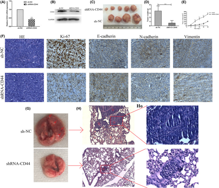
FIGURE 5.

Knockdown of CD44 inhibits in vivo tumorigenesis and metastasis of HCT116‐CSCs. (A) qPCR assay quantifies significantly lower CD44 expression in shRNA‐CD44‐transfected HCT116‐CSCs than in sh‐NC‐transfected HCT116‐CSCs; (B) Western blotting determines lower CD44 expression in shRNA‐CD44‐transfected HCT116‐CSCs than in sh‐NC‐transfected HCT116‐CSCs; (C) presence of xenograft tumors at the injection site 42 days postinjection; (D) the mean weight of the xenograft tumors derived from shRNA‐CD44‐transfected HCT116‐CSCs is significantly lower than from sh‐NC‐transfected HCT116‐CSCs; (E) the mean volume of the xenograft tumors derived from shRNA‐CD44‐transfected HCT116‐CSCs are all significantly smaller than from sh‐NC‐transfected HCT116‐CSCs 8, 16, 24, 32 and 40 days postinfection; (F) immunostaining analysis detects lower Ki‐67, N‐cadherin and vimentin expression and higher E‐cadherin expression in xenograft tumors derived from shRNA‐CD44‐transfected HCT116‐CSCs than from sh‐NC‐transfected HCT116‐CSCs; (G) representative images of nude mouse lung 6 weeks following injection of HCT116‐CSCs. Intrapulmonary metastatic nodules are seen in mice subcutaneously injected with sh‐NC‐transfected HCT116‐CSCs, while apparent intrapulmonary metastatic nodules are not visible in mice subcutaneously injected with shRNA‐CD44‐transfected HCT116‐CSCs. The red arrows indicate the intrapulmonary metastatic nodules; (H) HE staining displays multiple intrapulmonary metastatic nodules in mice subcutaneously injected with shRNA‐CD44‐transfected HCT116‐CSCs, while intrapulmonary metastatic nodules are visible in only one nude mouse subcutaneously injected with sh‐NC‐transfected HCT116‐CSCs. The red pentagrams indicate intrapulmonary metastatic nodules. Magnification, 100 ×; HO, Figure 5H at a magnification of 400×. **p < 0.01; ***p < 0.001
