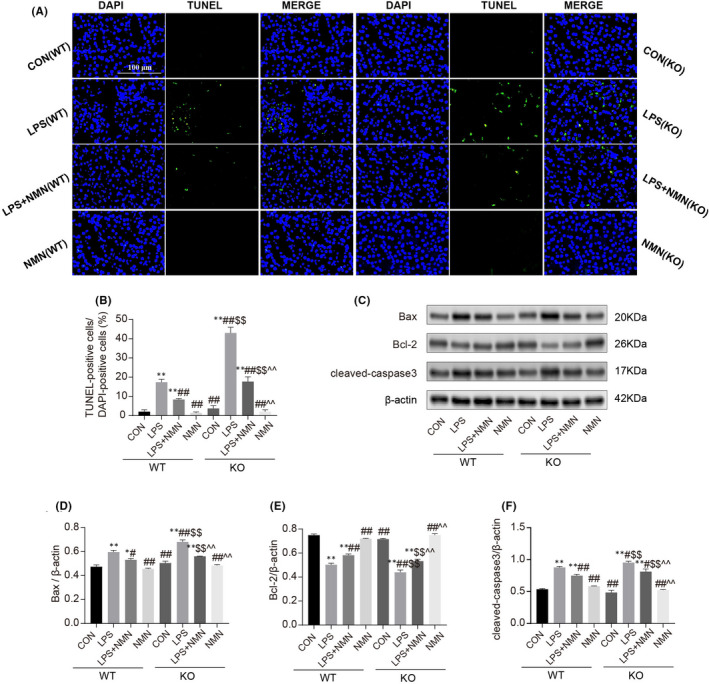
FIGURE 2.

Effect of NAD+ on LPS‐induced apoptosis in renal tissues. (A‐B) Representative images of the TUNEL staining on kidneys (scale bars, 100 μm) and counted percentages of terminal deoxynucleotidyltransferase‐mediated dUTP nick end‐labelling (TUNEL)‐positive cells. (C‐F) Representative immunoblotting analysis of Bax, Bcl‐2 and cleaved‐caspase3. Each result was replicated in three independent experiments, and all values are the mean ±SD (n = 3). *Significance compared with CON (WT) group (*p < 0.05, **p < 0.01). #Significance compared with LPS (WT) group (# p < 0.05, ## p < 0.01). $Significance compared with CON (KO) group ($ p < 0.05, $$ p < 0.01). ^Significance compared with LPS (KO) group (^ p < 0.05, ^^ p < 0.01)
