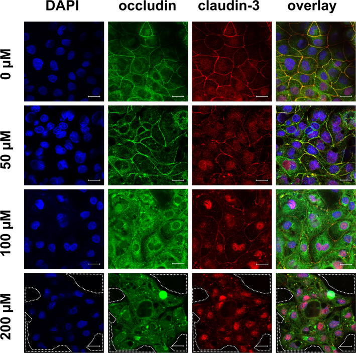
FIGURE 6.

Immunocytological staining of IPEC‐J2 cells on filter membranes with antibodies raised against occludin (green) and claudin‐3 (red) after 24 h of incubation with berberine. Nuclei were stained with DAPI (blue). Under control conditions, both TJ proteins could be located in the lateral membrane; the yellow signal in the overlay showed co‐localization. The signal for occludin appeared slightly stronger with 50 µM berberine, but 100 µM lead to a more intracellular signal, which was weaker in the 200 µM group. The claudin‐3 signal became weaker dose‐dependently. The area circled by white dots shows holes inside of the monolayer (scale bar: 20 µM, n = 4, representative images)
