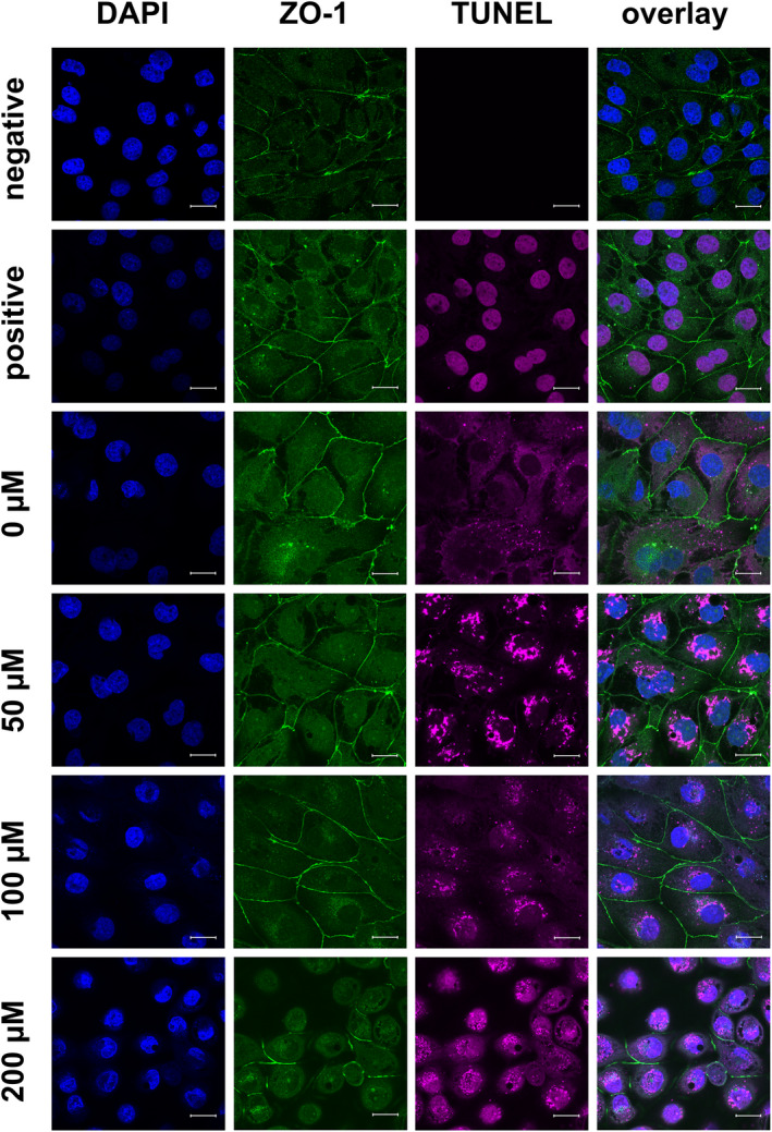
FIGURE 8.

TUNEL assay (purple) and immunocytological staining of IPEC‐J2 cells on coverslips with an antibody raised against ZO‐1 (green), Nuclei were stained with DAPI (blue). The cells used as TUNEL‐positive and ‐negative controls were treated as the control (0.2% DMSO). The negative control was incubated with blocking solution during the TUNEL assay, and the positive control was treated with DNAse to induce double‐strand breakage (seen as purple staining of the nuclei). As in Figure 5, the ZO‐1 signal was located near the lateral membrane and became weaker with increasing berberine concentrations. TUNEL staining was strongest following treatment with 50 µM berberine (scale bar: 20 µM, n = 3, representative images)
