Figure 6.

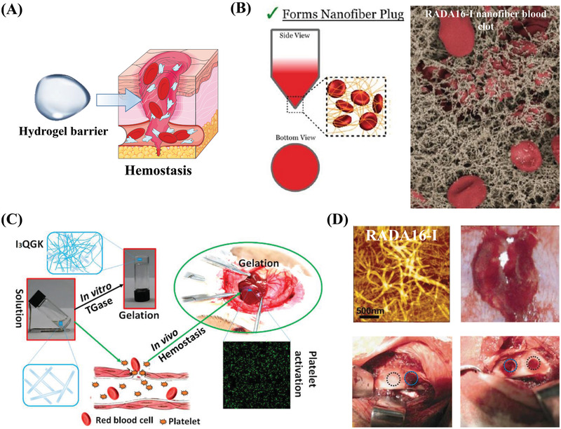
Hemostatic properties of self‐assembling peptides. A) Schematic illustration of self‐assembling peptide barriers for hemostasis. Part of the figure is modified from Servier Medical Art (http://smart.servier.com/), licensed under a Creative Common Attribution 3.0 Generic License. B) Hemostatic mechanism of a peptide nanofibrous hydrogel (RADA16‐I). Suspension assay shows that the nanofibers entangle with red blood cells to keep them in suspension, and the scanning electron microscopy (SEM) image shows that interwoven nanofibers entrap blood components and form clots to speed up hemostasis. Reproduced with permission.[ 86 ] Copyright 2015, American Chemical Society. C) Hemostatic properties and mechanism of a short‐peptide (I3QGK) hydrogel. I3QGK assembles into rigid hydrogels in the presence of transglutaminase (TGase) and shows adequate hemostasis by gelling blood and promoting platelet adhesion in a liver trauma model. Reproduced with permission.[ 88 ] Copyright 2016, American Chemical Society. D) Hemostatic properties of a longer peptide (RADA16‐I). AFM images show that the blood of the rabbit's middle auricular artery can induce the formation of a blood‐hydrogel. Hemostasis was achieved in 10 s (blue circles) in a cancellous ilium bone defect model. Reproduced with permission.[ 90 ] Copyright 2015, American Chemical Society.
