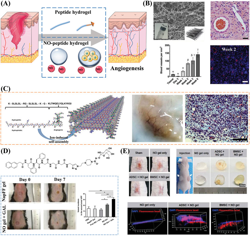
Figure 8.

Angiogenic properties of self‐assembling peptides. A) Schematic illustration of self‐assembling peptide and NO‐peptide hydrogels for promoting angiogenesis. Part of the figure is modified from Servier Medical Art (http://smart.servier.com/), licensed under a Creative Common Attribution 3.0 Generic License. B) The site of the subcutaneous injection of the peptide hydrogel K2(SL)6K2 is highly vascularized. The SEM image shows the fiber network structure formed by the self‐assembly of peptides. The H&E staining image of blood vessels in the hydrogel and plot of the number of blood vessels show that the implantation of the peptide hydrogel can promote angiogenesis. Reproduced with permission.[ 119 ] Copyright 2018, Elsevier Ltd. C) A VEGF‐binding self‐assembling peptide activates receptors to promote angiogenesis. After the subcutaneous implantation of the peptide hydrogel scaffolds, a large number of blood vessels can be seen. The Masson's Trichrome staining image further indicates the formation of blood vessels. Reproduced with permission.[ 120c ] Copyright 2015, American Chemical Society. D) NO‐releasing hydrogel promotes wound angiogenesis: β‐galactosidase triggers NO release and promotes peptide gelation; Number of stained micro‐vessels per HPF shows that NO gel + GAL group significantly promotes angiogenesis in wounded skin. Reproduced with permission.[ 122 ] Copyright 2013, Royal Society of Chemistry. E) NO‐gel and MSCs synergistically induce neovascularization. Images from in vivo wound healing experiments show that wound closure occurs faster in the BMSC‐embedded NO‐gel group compared to the other experimental groups. The gel plug angiogenesis assay results and 3D visualization of perfusable vessel formation indicate the ability of the NO‐gel to induce angiogenesis. Reproduced with permission.[ 127 ] Copyright 2020, AAAS.
