
Figure 4.
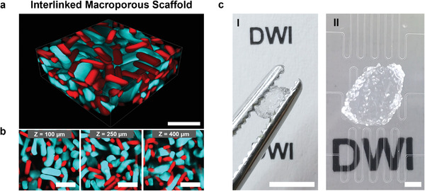
Interlinked microgel rod‐based scaffold. a) 3D projection of the 500 µm confocal microscopy Z‐stack of the interlinked scaffold made from epoxy‐functionalized microgel rods (7.90 mg mL−1 GMA, 10 wt% sPEG‐AC, red methacryloxyethyl thiocarbamoyl rhodamine‐B) mixed with the same number of amine‐functionalized microgel rods (12.15 mg mL−1 AMA, 10 wt% sPEG‐AC, cyan FITC). Scale bar represents 500 µm. b) Confocal Z‐stack images at different Z‐values (indicated in the insertion) representing the porous structure within the interlinked scaffold. Scale bars represent 500 µm. Movie S1 of the Supporting Information shows the Z‐stack. c) Images of an interlinked microgel rod‐based scaffold, between tweezers (I) and on top the employed microfluidic chips (II). Scale bars represent 5 mm in I and 1 mm in II.