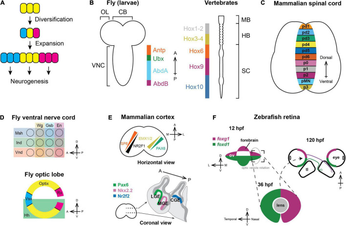
FIGURE 1.
Spatial patterning in neurogenesis. (A) General principles of spatial patterning in neurogenesis: Progenitors expand in number and then diversify the set of neuronal types that they can generate during neurogenesis, which is represented by different colors. (B) Nervous systems of vertebrates and invertebrates are patterned along the anteroposterior axis by Hox genes. Orthologous Hox genes have the same color (McGinnis and Krumlauf, 1992). (OL: optic lobe; CB: central brain; VNC: ventral nerve cord; MB: midbrain; HB; hindbrain; SC: spinal cord). (C) Vertebral neural tubes are patterned along the dorsoventral axis and form multiple progenitor domains that will generate spinal motor neurons and various interneurons (pd1-6: Progenitors for dorsal interneurons dI1-dI6; p0-3: Progenitors for ventral interneuron V0–V3; pMN: Spinal motor neuron progenitors). (D) Each segment of the fly ventral nerve cord (top) is partitioned along both the A–P and D–V axis by the expression of spatial factors; similarly, the Outer Proliferation Center neuroepithelium generating the medulla (bottom) is compartmentalized into six distinct domains. (E) Upper panel: In mammals, the cortical protomap is defined by the graded expression of transcription factors. SP8 and NR2F1 form opposing gradients where SP8 is highest on the anteromedial side while NR2F1 is highest on the posterolateral side. PAX6 and EMX1/2 form another set of opposing gradients along the anterolateral-posteromedial axis (Reviewed in Greig et al., 2013; Cadwell et al., 2019). Lower panel: Distinct subpallial progenitor domains are marked by specific transcription factors and generate different types of cortical inhibitory interneurons (LGE: Lateral ganglionic eminence; MGE: Medial ganglionic eminence; CGE: Caudal ganglionic eminence). (F) In zebrafish, the optic vesicle is dorsoventrally patterned by the expression of foxg1 and foxd1. During developmental eye rotation, the D–V axis becomes the nasotemporal axis, and the nasal hemiretina projects to posterior tectal lobe, while the temporal hemiretina projects to the anterior tectal lobe (hpf: hours post fertilization; ov: optic vesicle; tl: tectal lobe).

