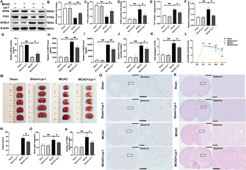
Fig. 2.
Suppression of ferroptosis and lipid peroxidation rescued cerebral I/R-induced brain injury in vivo. SD rats were treated with Lip-1 (i.p, 10 mg/kg) for 1 h and then subjected to MCAO operation. A–D The protein expression of GPX4, FTH1, and ASCL4 as assayed with western blotting. E, F Content of Fe2+ and total iron as determined with the iron assay kit. G GPX4 activity as detected using GPX4 activity assay kit. H, I The levels of 12-HETE and 15-HETE as determined with ELISA kits. J LPO level as assayed with the lipid peroxidation assay kit. K LDH level as quantified using LDH assay kit. L Neurological severity scores. M, N Infarct size in whole brain tissues as measured with TTC staining. O Brain water content. P TUNEL staining of rat brain tissues. Q HE staining of rat brain tissues (Scale bar = 50 μm). n = 6 rats/group, all data are expressed as the mean ± SD, *P < 0.05, **P < 0.01; MCAO group vs. the sham group and MCAO group vs. MCAO + Lip-1 group

