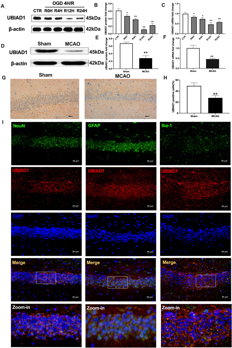
Fig. 4.
Expression and location of UBIAD1 in response to cerebral I/R injury in vivo and vitro. A–C The protein and mRNA expression of UBIAD1 in neurons as detected with western blotting and PCR assay. D–F The protein and mRNA expression of UBIAD1 in brain tissues as detected with western blotting and PCR assay. G, H Protein expression of UBIAD1 in brain tissues as evaluated through immunohistochemical staining (Scale bar = 50 μm). I Confocal images showing co-localization of UBIAD1 (red) with neuron marker NeuN (green), astrocytes marker GFAP (green) microglia marker Iba-1 (green) (Scale bar = 25 μm). n = 3 for in vitro, n = 6 rats/group for in vivo, all data are expressed as the mean ± SD, *P < 0.05, **P < 0.01; relative to the control group or sham group

