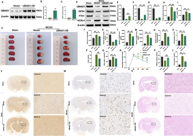
Fig. 7.
Overexpression of UBIAD1 suppressed brain injury induced by I/R-mediated lipid peroxidation and ferroptosis in vivo. A–C Overexpression of UBIAD1 by adeno-associated virus transfection as confirmed by western blotting and PCR assay in rats brain tissues. D–H The protein expression of UBIAD1, GPX4, FTH1, and ASCL4 in rat brain tissues after MCAO as determined by western blotting. I, J Content of Fe2+ and total iron in rat brain tissues after MCAO as evaluated using iron assay kit. K GPX4 activity in rat brain tissues after MCAO as assayed with the GPX4 activity assay kit. L, M 12-HETE and 15-HETE levels in rat brain tissues after MCAO as detected by ELISA assay kits. N LPO level in rat brain tissues after MCAO as determined with lipid peroxidation assay kit. O LDH level in rat brain tissues after MCAO determined by LDH assay kit. P and U Infarct size in whole rat brain tissues after MCAO as revealed by TTC staining. Q Brain water content after MCAO. R Confirmation of neurological severity scores after MCAO. S and V Protein level of UBIAD1 in rat brain tissues after MCAO as examined using immunohistochemical staining (Scale bar = 50 μm). T and W Neuronal death in rat brain tissues after MCAO as determined by the TUNEL staining assay (Scale bar = 50 μm). X HE staining in rat brain tissues after MCAO (Scale bar = 50 μm). n = 6 rats/group, all data are expressed as the mean ± SD, *P < 0.05, **P < 0.01; sham group vs. MCAO + vector-UBIAD1-AAV group and MCAO + vector-UBIAD1-AAV group vs. MCAO + UBIAD1-AAV group

