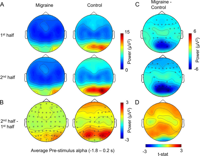
Fig. 6.
The average pre-stimulus alpha power change between 1st half and 2nd half of the experiment. A The voltage map showed that the pre-fixation alpha was the strongest at the occipital area. B Cluster-based permutation analyses displayed one significant cluster for migraine (p = .0002, t = − 1.8 to 0.25) and two for control (p = .0008, t = − 1.8 to − 0.75; p = .002, t = − 0.7 to 0.2). The significant channels are highlighted by an asterisk (*). C For the between group differences, there were one significant cluster for 1st half (p = .013) and one for 2nd half (p = .019) at the [− 1.8 to 0.2 s] interval, with the alpha power differences mainly distributed over the parietal-occipital region. D The pre-stimulus alpha power increase in the 2nd half was not significantly different between migraine and control groups

