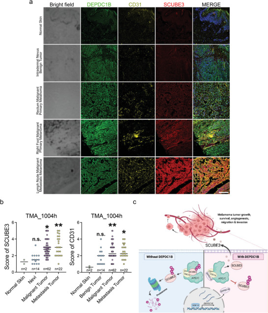
Figure 7.

SCUBE3 has a higher expression level in metastatic melanomas accompanied by denser blood vessel distribution. a) TMA immunofluorescence staining using anti‐SCUBE3 (red), anti‐DEPDC1B (green), and anti‐CD31 (yellow) together with DAPI (blue in the merge) for nuclei counterstaining. Scale bar = 50 µm. b) Quantitative analysis of the SCUBE3 protein level and micro vessel distribution (marked by CD31) in different stages of melanoma. c) Schematic diagram shows SOX10 directly transactivates DEPDC1B expression through binding to its promoter region. DEPDC1B competitively interacts with CDC16 in the cytosol to inhibit SCUBE3 ubiquitination. Stabilized SCUBE3 secreted from melanoma cells facilitates blood vessel recruitment to promote melanoma tumor growth, survival, and metastasis. Data are median with individual values. n.s. when p > 0.05; *p < 0.05; **p < 0.01; ***p < 0.001; ****p < 0.0001 from ordinary one‐way ANOVA.
