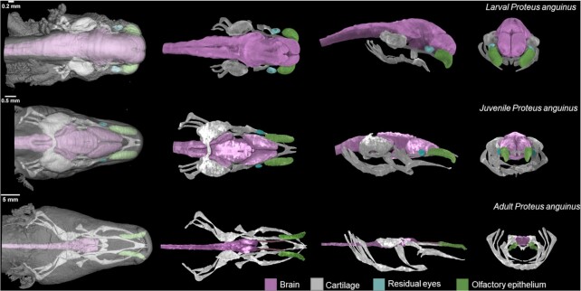
Figure 1:
3D reconstructions of P. anguinus head based on X-ray microCT data. Larva (top), juvenile (middle), and adult (bottom) P. anguinus. Images in the first column show semi-transparent 3D renderings of the head with skin in dorsal view. Dorsal, lateral, and frontal views of the segmented and color-coded internal soft structures are shown in the second to the fourth columns.

