
Figure 5.
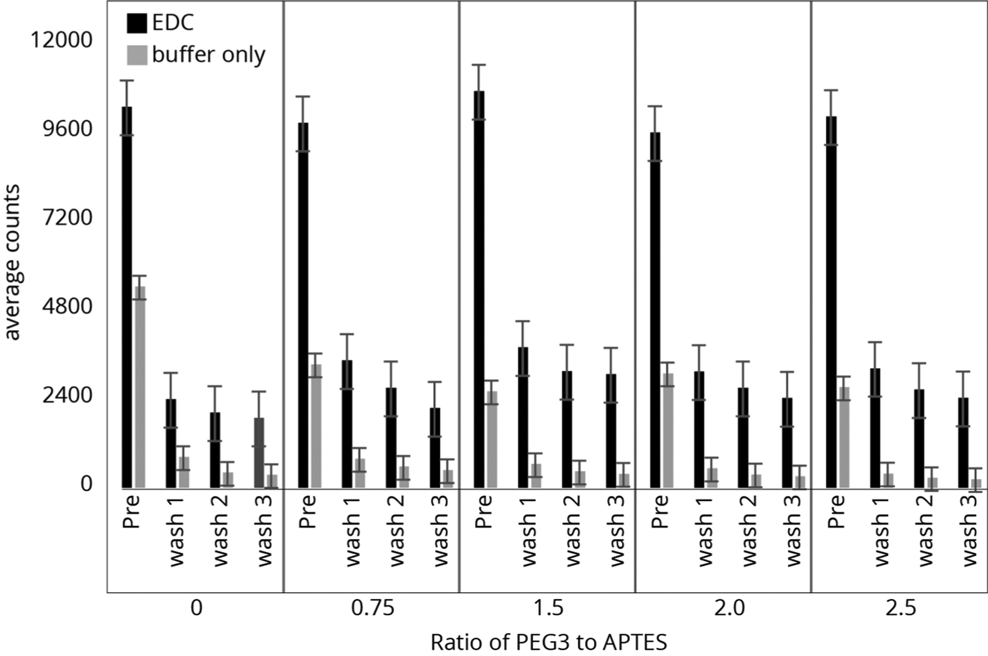
Counts of individual P1 peptide molecules from slide surfaces of APTES (0.43 mM) and PEG-3 (0–2.5 equiv). Atto647N-labeled peptides were bound to the surfaces before imaging and were washed with TFA three times, imaging after each cycle.
Official websites use .gov
A
.gov website belongs to an official
government organization in the United States.
Secure .gov websites use HTTPS
A lock (
) or https:// means you've safely
connected to the .gov website. Share sensitive
information only on official, secure websites.
Counts of individual P1 peptide molecules from slide surfaces of APTES (0.43 mM) and PEG-3 (0–2.5 equiv). Atto647N-labeled peptides were bound to the surfaces before imaging and were washed with TFA three times, imaging after each cycle.