. 2022 Mar 8;12(13):7635–7651. doi: 10.1039/d2ra00566b

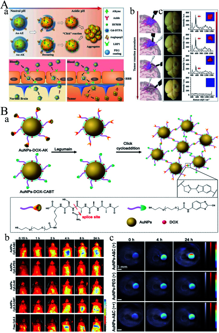
Fig. 5. (A) Guiding brain-tumor surgery by acid-responsive gold nanoprobes. (a) The cleavage of the PEG coating in physiological acidity triggers aggregation between Au-AZ and Au-AK via “click” cycloaddition reactions. (b) Au-AZ/Au-AK mixture intraoperatively guiding glioma resection using a handheld Raman detector. Cartoons illustrating the procedure of Raman-spectroscopy-guided glioma resection. (c) Photographs of the excised mouse brain bearing U87MG glioma xenografts and Raman spectra at the tumor site during the image-guided tumor resection. Surgery was conducted at 24 h PI of the Au-AZ/Au-AK mixture (1 : 1 molar ratio, totaling 600 pmol kg−1). The intensities of the characteristic Raman twin peak were quantified in a 5 × 5 grid covering the tumor cutting bed. Reproduced with permission from ref. 37, copyright: 2017, Wiley. (B) (a) Diagram depicting the legumain-triggered aggregation and composition of AuNPs-DOX-A&C. The AuNPs-DOX-AK undergoes a cleavage by legumain to expose 1,2-thiol amino group of cysteine, where click cycloaddition further occurs with the contiguous cyano group of AuNPs-DOX-CABT to form aggregates. (b) Living imaging of BALB/c mice after intravenous injection with AuNPs-Cy5.5-A&C and control formulations for different time intervals; bar indicates radiant efficiency from 0.5 × 10⁷ to 2.5 × 10⁷. (c) PA images of AuNPs-A&C incubated with legumain in the absence and presence of Ato acquired from phantom at 690 nm, and AuNPs-PEG incubated with legumain; gray bars indicate ultrasound image of phantom, red bars indicate PA signal of phantom, and jet bars indicate the PA signal of AuNPs. Reproduced with permission from ref. 38, copyright: 2016, American Chemical Society.

Fig. 5