. 2022 Mar 8;12(13):7635–7651. doi: 10.1039/d2ra00566b

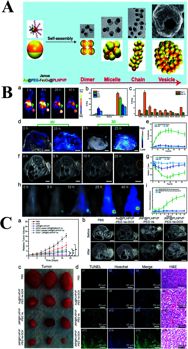
Fig. 8. (A) Schematic representation of the self-assembly of amphiphilic Janus NPs into dimer, micelle, chain, and bilayered vesicles and their corresponding simulation results. (B) In vivo imaging and biodistribution studies of the hybrid vesicles. (a) PET imaging of U87MG tumor-bearing mice at different time points after intravenous injection of [64Cu] radiolabeled JNP vesicles. (b) PET region of interest (ROI) analysis of tumor, muscle, and liver at 1, 5, 25, and 40 h postinjection. (c) Direct tissue sampling biodistribution in different organs at day 1 and 12 postinjection analyzed by ICP-MS. (d) In vivo PA images and (e) average PA intensity in the tumor after intravenous injection of the hybrid vesicles. (f) T2-weighted MR images and (g) the relative T2 values of the tumor before and after intravenous injection of vesicles at different times. Scale bar: 5 mm. (h) In vivo fluorescence imaging and (i) tumor signal intensity after injection of IRDy800CW-loaded vesicles. (C) In vivo cancer therapy effect of JNP vesicles. (a) Tumor growth profiles in mice after different treatments (*P < 0.05; **P < 0.01). (b) Tumor MR images before and 18 days after treatment with different formulations. (c) Representative photographs of the dissected tumors in different treatment groups. (d) TUNEL-labeled tumor sections treated with different formulations (green color: positive TUNEL staining, blue color: nuclei stained by Hoechst 33342) and histological analysis of tumor sections after staining with H&E in different treatment groups. Reproduced with permission from ref. 59, copyright: 2019, ACS.

Fig. 8