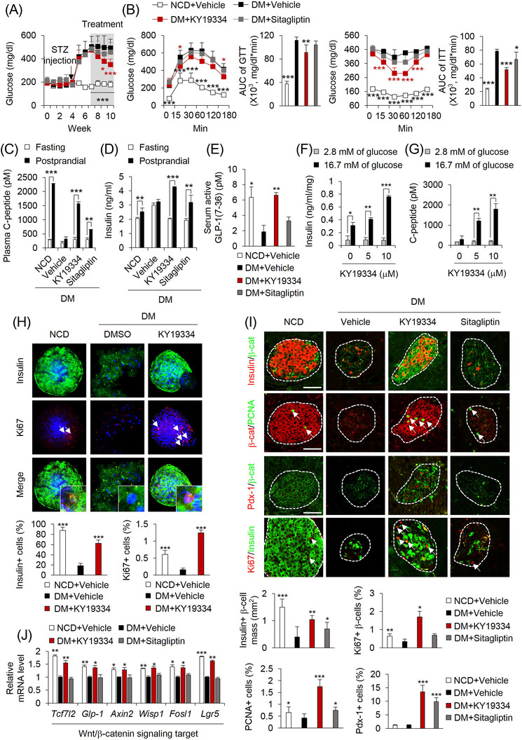
FIGURE 6.

KY19334 treatment restores β‐cell mass and functions in HFD‐fed and STZ‐induced diabetes mellitus (DM) mice. C57BL/6 mice fed NCD or HFD for 4 weeks followed by injection with STZ (50 mg/kg/d) for 1 week. Afterward, mice were p.o. administered KY19334 (25 mg/kg/d) or sitagliptin (50 mg/kg/d) for 4 weeks (n = 6 per group). (A) Non‐fasting blood glucose levels. (B) Glucose tolerance and insulin tolerance test and AUC. (C–E) The concentration of plasma C‐peptide (C), insulin (D) and serum active GLP‐1 (E). (F–H) Isolated islets from the pancreas of NCD or DM mice treated with DMSO or KY19334 for 72 h. The concentration of secreted insulin (F) and C‐peptide (G) from islets in response to different concentrations measured after incubation for 1 h with either low (2.8 mM) or high (16.7 mM) glucose in KRBH buffer. (H) Representative images of immunofluorescent staining for insulin and Ki67 (upper panel). Quantitative analyses of insulin‐ and Ki67‐positive cells, respectively, in the islets (lower panel). (I) Representative images of IHC staining of the pancreas for insulin, β‐catenin, PCNA, Pdx‐1 and Ki67. Arrows indicate proliferating β‐cells (upper panel). Quantitative analyses of insulin‐positive β‐cell mass, PCNA, Pdx‐1 and Ki67 positive cells in pancreatic tissues (lower panel). (J) Relative mRNA expression of Wnt/β‐catenin signalling target genes. Expression levels of mRNA were normalized by vehicle‐treated HFD mice group. Scale bars = 100 μm. All data are presented as the mean ± SD. *p < 0.05, **p < 0.01, ***p < 0.001 determined by Student's t‐test. DM: Diabetes mellitus; β‐cat: β‐catenin
