Figure 3.
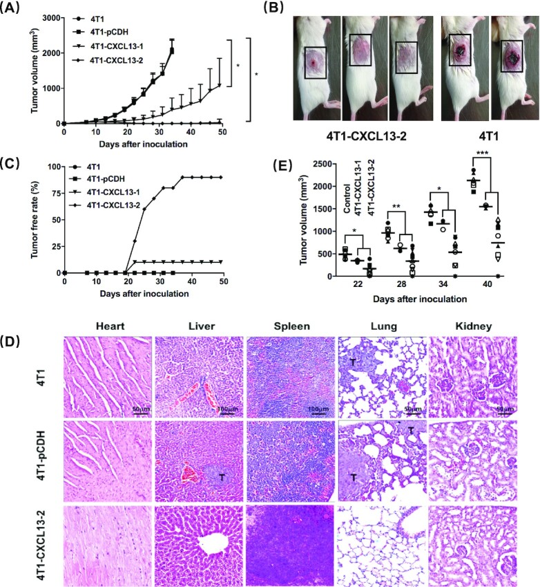
CXCL13 expression in 4T1 tumor microenvironment impaired the in vivo tumor growth and metastasis. (A) BALB/c mice were injected s.c. with 2 × 10⁵ 4T1, 4T1-pCDH, 4T1-CXCL13-1, or 4T1-CXCL13-2 cells, and tumors were calculated every 3 days. Bars, means ± SD (n = 10). This experiment was repeated 3 times (*P < 0.05). (B) Representative pictures of mice after 21 days of 4T1-CXCL13-2 (left) or parental 4T1 (right) inoculation; tumor site is shown in the black-dotted rectangle. (C) Tumor-free rate curve of each group. (D) Tumor metastases were observed in major organs by H&E pathological staining (T = tumor; lung/heart/kidney magnification, ×200; liver/spleen magnification, ×100). (E) Mice from 4T1-CXCL13-1 (n = 3) and 4T1-CXCL13-2 (n = 9) groups with >6 months complete tumor regression were rechallenged with parental 4T1 cells (2 × 10⁵), and tumor growth was significantly inhibited in both groups. The control group was naïve BALB/c mice inoculated with 2 × 10⁵ 4T1 cells (n = 6) (*P < 0.05, **P < 0.01, ***P < 0.001).

