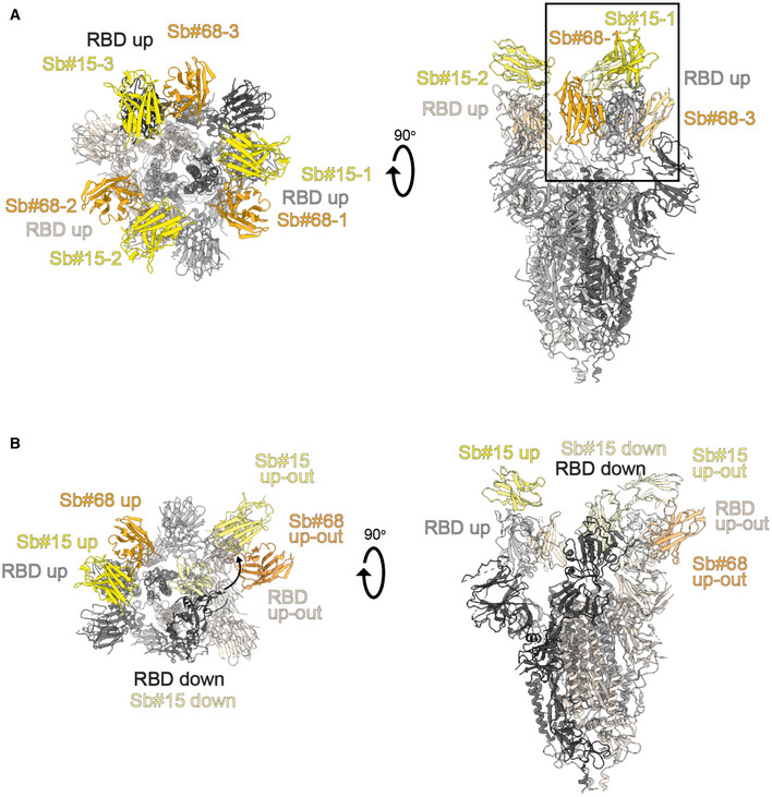
Figure EV2. Structures of S‐2P spike in complex with both Sb#15 and Sb#68.

- Structure of S‐2P with both Sb#15 and Sb#68 bound to each RBD adopting a symmetrical 3up conformation. Based on the cryo‐EM map shown in main Fig 5A, a model shown as ribbon was built using pre‐existing structures (PDB ID:6X2B for S‐2P; PDB ID:3K1K for Sb#15; PDB ID:5M13 for Sb#68).
- Structure of S‐2P with the three RBDs adopting an asymmetrical 1up/1up‐out/1down conformation. Based on the cryo‐EM map shown in Fig 5B, a model shown as ribbon was built using pre‐existing structures (PDB ID:6X2B for S‐2P; PDB ID:3K1K for Sb#15; PDB ID:5M13 for Sb#68). The up‐out state is pushed outward by the adjacent RBD in a down state with bound Sb#15 (arrow). Spike protein is shown in shades of gray, Sb#15 in yellow, and Sb#68 in orange.
