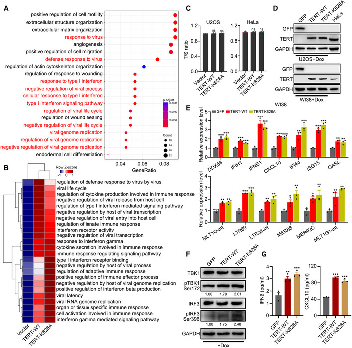
Figure EV1. Ectopic expression of TERT triggers interferon response.

-
AEnriched biological processes (BP) with TERT ectopic expression. GO was analysed for upregulated genes by TERT compared to those in the empty vector control. GO terms in red are biological processes related to interferon response.
-
BssGSEA of GO terms related to interferon response in U2OS cells ectopically expressing TERT‐WT or TERT‐K626A.
-
CT/S ratio of U2OS and HeLa cells with transient transfection of TERT‐WT or TERT‐K626A.
-
DWestern blot analysis of GFP, TERT and GAPDH levels in U2OS (upper) and WI38 (lower) cells ectopically expressing TERT‐WT or TERT‐K626A using Dox‐inducible system to induce expression of TERT at the levels comparable to that of HeLa cells. GFP as negative control.
-
E–GRT–qPCR analysis of interferon‐related genes and TA‐ERVs (E), western blot analysis of TERT, TBK1, pTBK1, IRF3, pIRF3, and GAPDH levels (F), and ELISA of IFNβ and CXCL10 in culture supernatant (G) in WI38 cells ectopically expressing TERT‐WT or TERT‐K626A using Dox‐inducible system. GFP as negative control. Relative pTBK1 and pIRF3 protein levels were quantified with ImageJ software and normalized to GAPDH, as indicated at the bottom of the blot (F).
Data information: Data represent mean ± SEM of three biological replicates (C, E and G). *P < 0.05, **P < 0.01, ***P < 0.001, ns, not significant, using one‐way ANOVA with Fisher's LSD tests (C, E and G).
Source data are available online for this figure.
