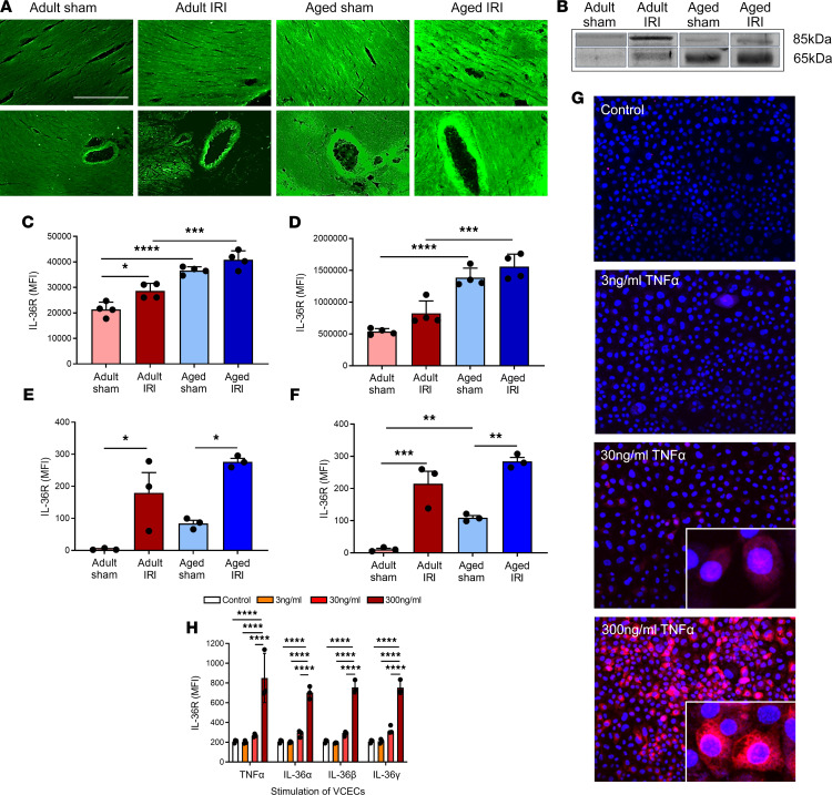
Figure 1. Age increases expression of IL-36R in healthy and IR-injured hearts, and expression can also be induced in endothelial cells with TNF-α or IL-36 cytokine stimulation.
Hearts were assessed for IL-36R using immunostaining, Western blot analysis, and flow cytometry. Representative images of IL-36R (green) staining of (A) frozen heart sections and (B) Western blots. The molecular weight of IL-36R is about 65 kDa, but it migrates to the position of about 85 kDa in denaturing protein gels. Hence, 2 bands were observed corresponding to 65 kDa, the more active protein due to cleavage of its signaling peptide, and 85 kDa, the less potent glycosylated form. Quantitative analysis of the (C) immunofluorescence images (n = 4/group) and (D) Western blots (n = 4/group). P = 0.0146 adult sham vs. adult IR immunofluorescence. P = 0.0003 aged sham vs. aged IR immunofluorescence. P = 0.001 aged sham vs. aged IR Western blot. Hearts were also collagenase digested and analyzed flow cytometrically for IL-36R expression. Flow cytometry analysis confirmed that IR injury and age induced a significant increase in IL-36R on (E) coronary endothelial cells and (F) cardiomyocytes (n = 3/group). (G and H) Murine vena cava endothelial cells (VCECs) were cultured and stimulated for 4 hours with experimental media (control), an IL-36 cytokine (α, β, or γ), or TNF-α. (G) Representative images of IL-36R (red) expression on stimulated, nonpermeabilized cells (Hoechst 33342–stained nuclei in blue). (H) Quantitative analysis of IL-36R expression on VCECs following stimulation (n = 3/group). Scale bar indicates 200 μm. *P < 0.05, **P < 0.01, ***P < 0.001, ****P < 0.0001 as determined using a 1-way ANOVA followed by a Tukey’s post hoc test.

