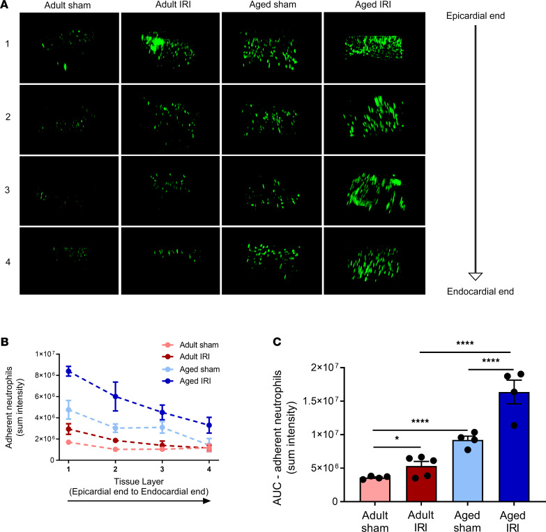
Figure 7. Age increases neutrophil presence within the deeper layers of the healthy and IR-injured myocardium.
(A) The left ventricle was vibratome sectioned into four 300 μm sections and imaged using a multiphoton microscope. Representative Z-stack multiphoton images of neutrophils (green) in the 4 layers of the left ventricle taken from the outermost layer closest to the epicardium (first row), outer myocardial layer (second row), inner myocardial layer (third row), and innermost layer closest to the endocardium (fourth row). Quantitative analysis of the multiphoton data at various depths for (B) adherent neutrophils and corresponding (C) AUC for adherent neutrophils. Adult sham — n = 4/group; adult IRI — n = 5/group; aged sham — n = 4/group; aged IRI — n = 4/group. *P = 0.0325, ****P < 0.0001 as determined using a 1-way ANOVA followed by a Tukey’s post hoc test.

