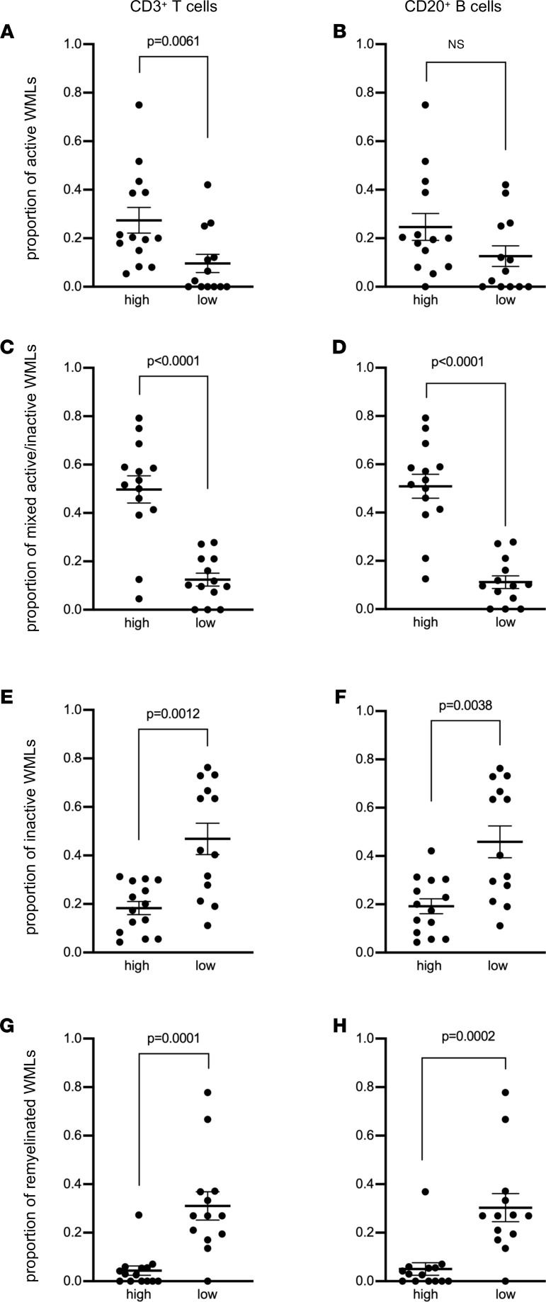
Figure 2. Enrichment of meningeal T cells and B cells is linked to subcortical white matter lesion activity.
Quantification of (A and B) the proportion of active white matter lesions (WMLs), (C and D) mixed active/inactive WMLs, (E and F) inactive WMLs, and (G and H) remyelinated WMLs in MS donors with high (n = 14) versus low (n = 13) meningeal CD3+ T cells (A, C, E, and G) or CD20+ B cell (B, D, F, and H) count. Each data point represents the proportion of WMLs in all tissue blocks analyzed per case (range 5–72 blocks, median: 30 blocks per donor). Statistically significant differences were determined by the nonparametric Mann-Whitney test.

