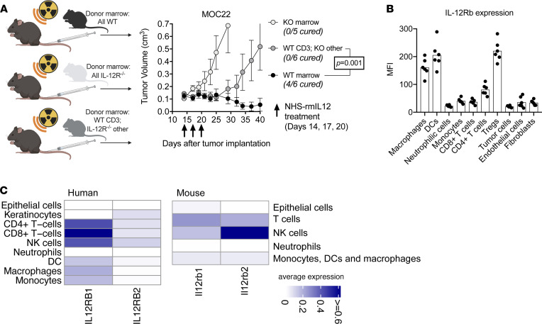
Figure 7. Direct effects of NHS–rmIL-12 on the lymphoid and myeloid compartment are necessary for tumor cure.
(A) Schematic of chimera experiments (left panel). WT B6 mice were irradiated to 9 Gy and transplanted with WT marrow, IL-12Rb2–KO marrow, or mixed marrow consisting of WT CD3+ marrow cells and IL-12Rb2–KO non-CD3+ marrow cells (n = 5–6/group). Three weeks after transplantation, all mice were treated with 3 low-dose NHS–rmIL-12 treatments and followed for tumor growth. Significance determined by 2-way ANOVA. (B) Day 10 MOC22 tumors (n = 6) were dissociated, and the MFI of IL-12Rb2 expression was quantified on individual cell types via flow cytometry. (C) Heatmaps show average normalized expression of IL-12R subunits in different cell subsets identified in single-cell RNA-Seq data of 18 human papillomavirus–negative head and neck SCCs and 2 control-treated MOC22. Of note, only a few cells were classified as epithelial or keratinocyte in human single-cell RNA-Seq data (about 10 cells), as these experiments were performed using sorted immune cells.

