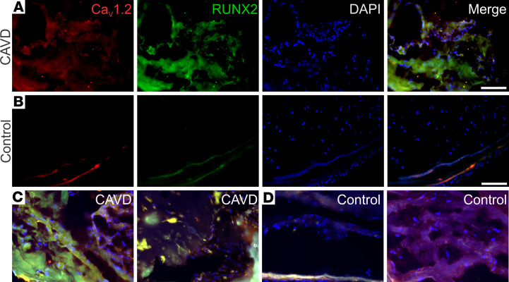
Figure 1. Increased CaV1.2 and RUNX2 expression in calcified segments of aortic valves from individuals with CAVD.
(A and B) IHC analysis of CaV1.2 (red) and RUNX2 (green) of valve tissue excised from a patient with CAVD (A) or a valve excised from a heart harvested for transplant and without CAVD (B). The blue signal is DAPI, and merged images are shown in the right-most panels for each. (C and D) Merged images for 2 additional valves from patients with CAVD (C) and from valves excised from hearts harvested for transplant and without CAVD (D). Scale bars: 100 μm.

