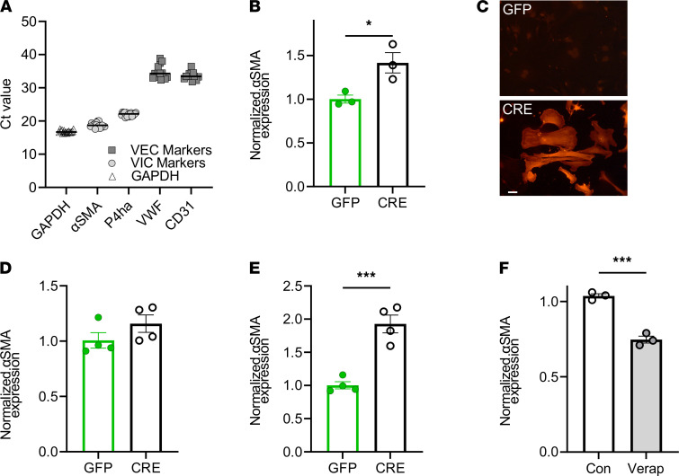
Figure 5. Blockade of Ca2+ influx through CaV1.2 reduces aortic valve lesions in Scx-CaV1.2TS mice.
(A) qPCR showing expression of VIC markers (α-SMA and P4ha) compared with endothelial cell markers (VWF and CD31) in cultured VICs. (B) Normalized (to GAPDH) α-SMA expression after 48 hours in CaV1.2WT-expressing VICs (after infection with virus expression GFP or Cre recombinase). *P < 0.05. (C) Anti–α-SMA immunofluorescence in CaV1.2TS VIC cultures expressing Cre or GFP. Scale bar: 50 μm. (D) Normalized (to GAPDH) α-SMA expression after 48 hours in WT VICs (after infection with virus expression GFP or Cre recombinase). P = NS. (E) Normalized (to GAPDH) α-SMA expression after 48 hours in CaV1.2TS expressing VICs (after infection with virus expression GFP or Cre recombinase). P < 0.001. (F) qPCR shows verapamil decreases relative expression of α-SMA in CaV1.2TS expressing VICs. ***P < 0.001. (B–F) Statistical comparisons were performed with 2-sided t tests. Con, control; VEC, valve endothelial cell.

