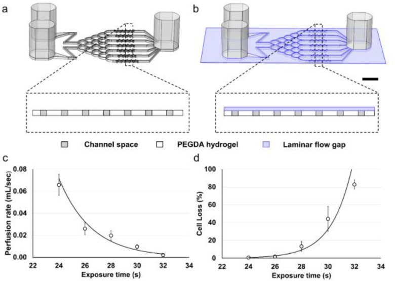
Figure 1.
Schematic and optimization of the brain cancer chip and microfluidics application using a polyethylene glycol diacrylate (PEGDA) hydrogel. (a) Schematic of the original brain cancer chip design, without the laminar flow layer, and a cross section over the microwell array. (b) Schematic of the brain cancer chip design with the additional laminar flow layer, and a cross section over the microwell array. Scale bar is 5 mm. (c) Perfusion decreases as exposure time increases (n = 4). (d) Cell loss increases during the cell seeding stage as exposure time increases (n = 4).

