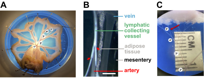
Figure 4. Isolation of small mesenteric arteries for vascular studies.
A. The isolated rat mesenteric bed is pinned out in a Petri dish containing colored silicon elastometer and filled with physiological salt solution. B. Magnified brightfield image showing distinctions between different classes of vessels and tissue in the mesentery. Scale bar: 1 mm. C. Magnified image illustrating the 5th order artery selected for cannulation after removal of the adipose tissue, veins, lymphatic vessels, and connective tissue. Figure 3A and 3C were provided by Perenkita Mendiola at the Dept. of Cell Biology and Physiology, Univ. of New Mexico, Albuquerque, NM, and used with permission ( Wenceslau et al., 2021 ). Figure 3B is used with permission from Sabine et al. (2018) .

