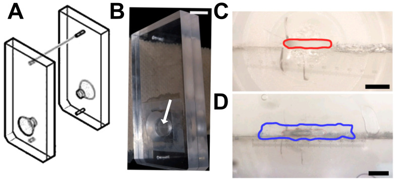
Figure 6. Tissue cassette designed for vessel analysis.
A. Diagram of a tissue cassette, used with permission from Warner Instruments, ©2022. The sample is placed within the two halves of the cassette. B. Arrow indicates the horizontal opening of the cassette in the photograph. Scale bar, 1 cm. C–D. Magnified image of the cassette showing the opening with an approximate area of 0.26 mm2 (red line; C), which can be completely covered with a longitudinally sectioned vessel (border of the vein marked in blue; D). Scale bars, 750 μm (C) and 650 μm (D). Numerical marks in C and D reflect eyepiece magnification. Figures B-D are from Maier-Begandt et al. (2021) , used with permission.

