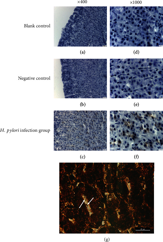
Figure 4.

Immunohistogram of H. pylori in gastric mucosa. (a) Blank control, ×400; (b) negative control, ×400; (c) experimental group, ×400; (d) blank control, ×1000; (e) negative control, ×1000; and (f) experimental group, ×1000. (g) Warthin-Starry silver staining on gastric mucosa of H. pylori infected gerbil, ×1000.
