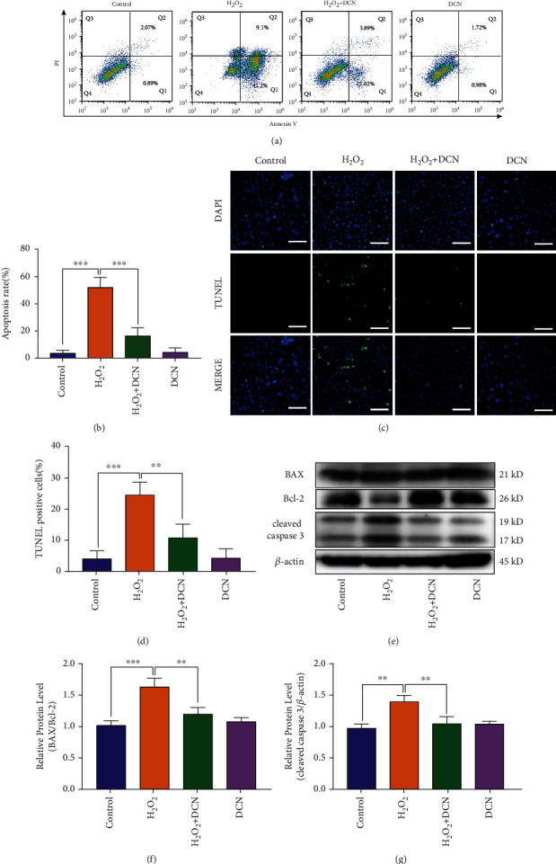
Figure 3.

DCN ameliorated the cell apoptosis in oxidative stress. (a) Representative flow cytometry plots using Annexin V-FITC/PI staining for apoptosis in ARPE-19 cells. (b) Quantification of apoptotic cells. (c) TUNEL staining was applied to measure apoptosis levels in ARPE-19 cells. (d) Quantification of TUNEL staining. (e–g) Western blot analysis and quantitative analysis of BAX, BCL2, and Cleaved-Caspase 3 protein level in ARPE-19 cells. Data is shown as mean ± SD, ∗p < 0.05, ∗∗p < 0.01, and ∗∗∗p < 0.001.
