
Figure 3.
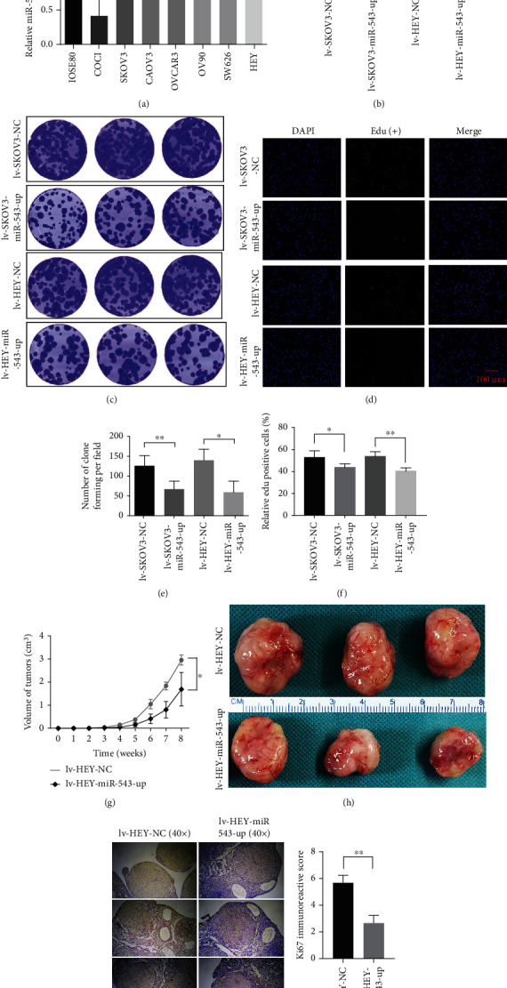
In vitro and in vivo functional assays of miR-543 in OvCa. (a) Relative miR-543 quantitative expression in normal ovarian cell lines and wild-type OvCa cell lines. (b) The stable upregulated expression of miR-543 was tested in SKOV3 and HEY cells by RT-PCR. Then, the stable clone forming (c, e) and Edu (d, f) assays revealed a significantly decreased proliferation ability after stable overexpression of miR-543. (g, h) In vivo assays showed that the tumour volume of the OvCa subcutaneous xenograft of lv-HEY-miR-543-up cells was significantly lower than that of the control cells. (i, j) Ki67 staining was significantly decreased in the subcutaneous xenograft of lv-HEY-miR-543-up cells compared with that in controls. Abbreviation: lv: lentivirus; Edu: 5-ethynyl-2′-deoxyuridine. ∗P < 0.05 vs. control (unpaired t-test), ∗∗P < 0.01 vs. control (unpaired t-test), and ∗∗∗P < 0.001 vs. control (unpaired t-test).