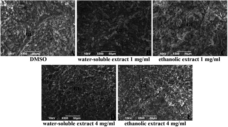
Fig. 4.
Representative scanning electron microscopy (SEM) images of metacestodes (MTCs) after water-soluble and ethanolic extracts of Capparis spinosa fruit incubation for 11 days. (A) MTCs incubated in complete culture medium containing 1% dimethyl sulfoxide (DMSO) served as a solvent group; (B) MTCs incubated with 1 mg/ml water-soluble extracts of C. spinosa fruit; (C) MTCs incubated with 1 mg/ml ethanolic extracts of C. spinosa fruit; (D) MTCs incubated with 4 mg/ml water-soluble extracts of C. spinosa fruit; (E) MTCs incubated with 4 mg/ml ethanolic extracts of C. spinosa fruit. GL, germinal layer.

