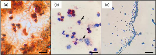
Fig. 1.

Microscopic images of gram-stained unconcentrated milk cells without centrifugation (a), concentrated milk cells with centrifugation (b), and isolates from the same mastitic milk sample (c). Arrows in (b) indicate the causal mastitis bacteria in milk. Scale bars=20 μm.
