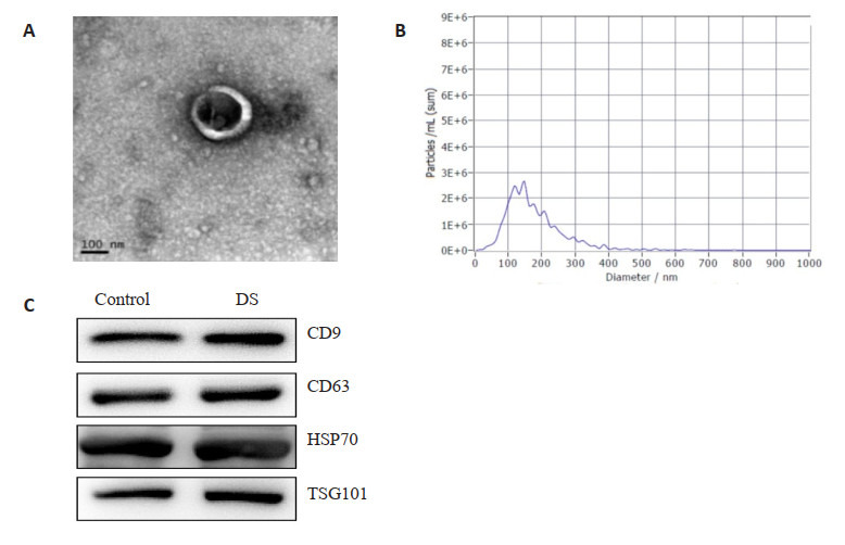
图 1.

羊水外泌体鉴定
Identification of exosomes in amniotic fluid. A: Exosome morphology under transmission electron microscope (arrow). B: Particle size of the exosomes. C: Expressions of exosome markers CD9, CD63, HSP70 and TSG101 detected with Western blotting.
