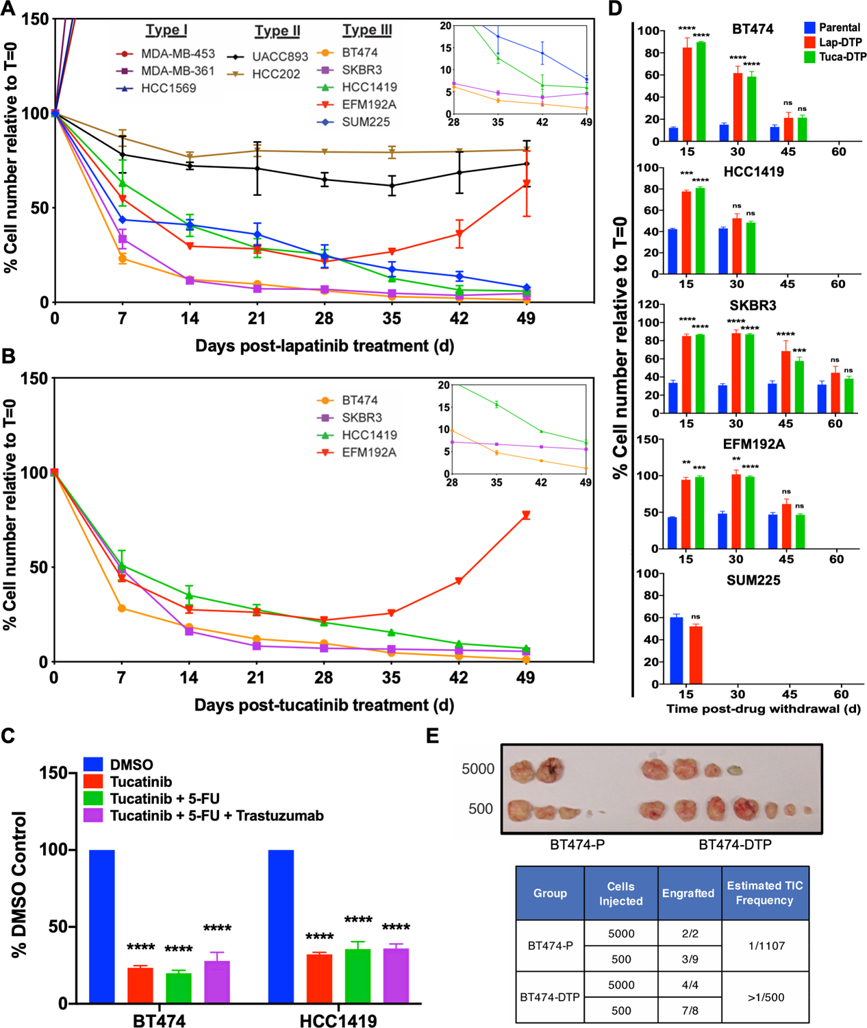
Figure 1. Some HER2+ breast cancer cell lines give rise to HER2 TKI drug-tolerant persisters (DTP).

A and B, HER2+ breast cancer cell lines were treated with 2.5 μM lapatinib (A) or with 1.2 μM tucatinib (B), counted at the indicated times, and the percentage of the initial cell number was determined. Mean ± SEM from three independent experiments is displayed shown. The inset shows a magnified view of the residual cells from Days 28–49. C, BT474 and HCC1419 cells were treated with the indicated agents for 14 days and percentage survival was quantified. D, Cells from the indicated lines were cultured in lapatinib or tucatinib (as indicated) for 14 days to generate DTPs. Then drug was withdrawn, and cells were allowed to resume proliferation. At the indicated times, cells were re-challenged with the same drug. Parental cells were used as a control at each time point. Cell survival was assessed by counting viable cells after 7-days of TKI treatment and normalized to the viable cell number at Day 0 (t=0). Mean ± SEM from three independent experiments is displayed. Statistical significance was assessed by two-tailed t-test for SUM225. All other cell lines were assessed by two-way ANOVA with Bonferroni post-hoc analysis (ns, p>0.05, *p<0.05 **p<0.01, ***p<0.001, ****p<0.0001). E, Number of tumors detected and estimated tumor-initiating cell (TIC) frequency of BT474 parental cells and lapatinib-DTPs after injection into the mammary fat pad of NSG mice. The top image shows tumors 5 months post-injection.
