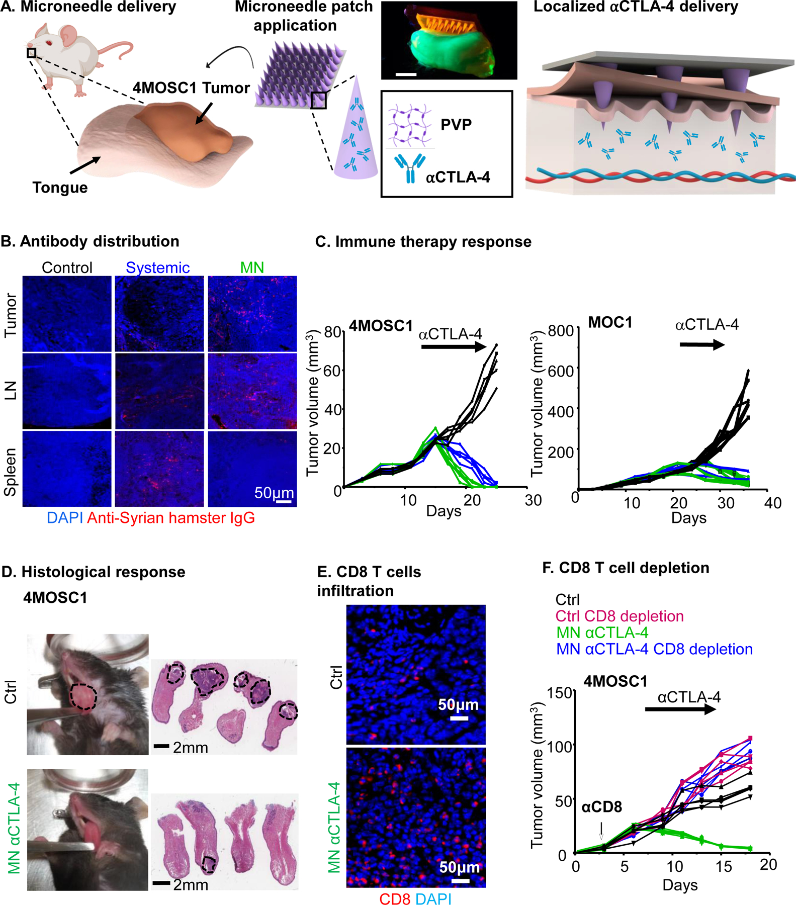
Figure 1.

A) Schematic of a dissolvable MN application on 4MOSC1 tumors in the tongue of mice for the delivery and release of an IO agent (⍺CTLA-4) to treat HNSCC. Digital photograph of the application of a dye supplemented (Rh6G) MN patch onto a fluorescent synthetic FITC hydrogel mouse tongue. Scale bar, 3mm. B-D) C57Bl/6 mice were implanted with 1 × 10⁶ 4MOSC1 cells into the tongue. After the tumors reached ~30 mm3, mice were treated with PVP patches (MN, control), or αCTLA-4 systemic (5 mg/kg) or by MN (0.1 mg) delivery. B). Shown is the immunofluorescent staining of the distribution of anti-Syrian hamster IgG (isotype for αCTLA-4 antibody) in mice with 4MOSC1 tumors treated by negative MN (control), αCTLA-4 s (IP, systemic) or αCTLA-4 MN. Staining for anti-hamster IgG (red) showed the localization of αCTLA-4 antibody in the tongue, lymph nodes, and spleen of treated mice. DAPI staining for nuclei is shown in blue (representative from 3 independent experiments with each n = 3 mice per group). C) Individual growth curves of 4MOSC1 and MOC1 tumor-bearing mice. Primary tumor growths are shown (n = 5 mice per group) using control (black), αCTLA-4 IP (blue), or αCTLA-4 MN (green). D) Histological responses. Left panel, representative pictures of tongues from isotype control and αCTLA-4 MN groups. Middle panel, representative H&E staining of histological tissue sections from mouse tongues from isotype control and αCTLA-4 MN groups. Sale bars represent 2mm. E) Immunofluorescence staining showing increased CD8 T-cells recruitment in the tumor due to MN αCTLA-4 treatment. Scale bars represent 50μm. F) Dependency of MN αCTLA-4 on CD8 T-cells. C57Bl/6 mice were treated with CD8 T-cell depleting antibody daily for 3 days before tumor implantation and then once a week after. HNSCC tumor bearing mice (as above) were treated with negative PVP MN (control) or αCTLA-4 MN (twice a week) (n = 5 per group). Individual growth curves of 4MOSC1 tumor-bearing mice are shown.
