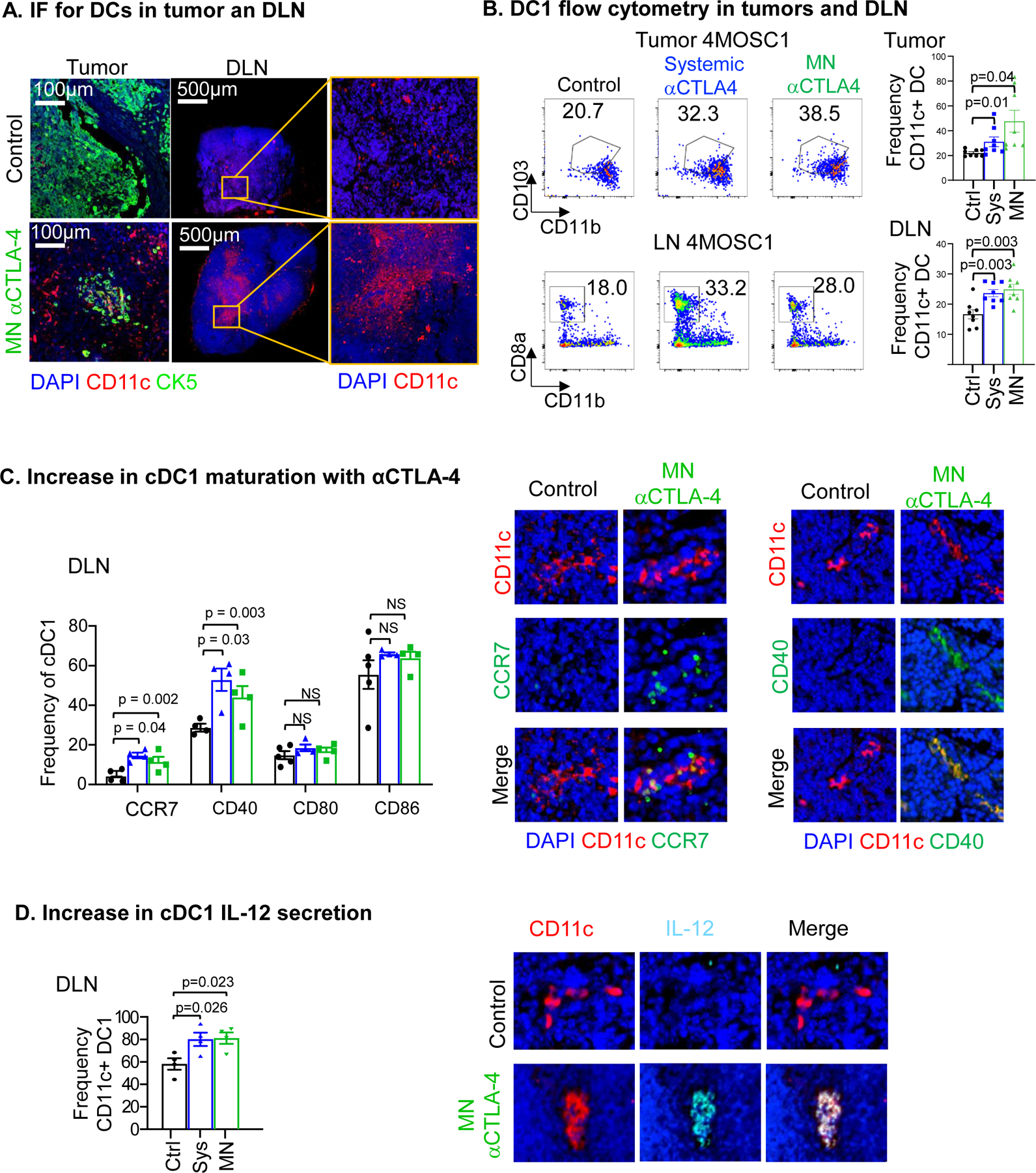
Figure 2.

A) Immunofluorescence staining of CD11c (red) in tumors and cervical draining lymph nodes (DLN) highlights an increase in CD11c+ dendritic cells recruitment with αCTLA-4 treatment. Immunofluorescence staining of CK5 (green) show squamous cell character of the lesion and DAPI in blue. B) Flow cytometry analysis showing cDC1s increase intratumorally and in DLN with αCTLA-4 treatment comparing systemic and MN delivery strategies (n = 8 mice per group). C) Increase in cDC1 maturation markers with αCTLA-4 in the lymphatic compartment by flow cytometry (left, n = 3–5 mice per group) and representative IF staining (right). D) αCTLA-4-mediated increase of IL-12 secretion by cDC1s in the lymph node by flow cytometry (n = 3–5 mice per group) and representative IF stainings (right). Data are reported as mean ± SEM; two-sided Student’s t-test; the p-value is indicated where relevant when compared with the control-treated group; non-significant (ns).
