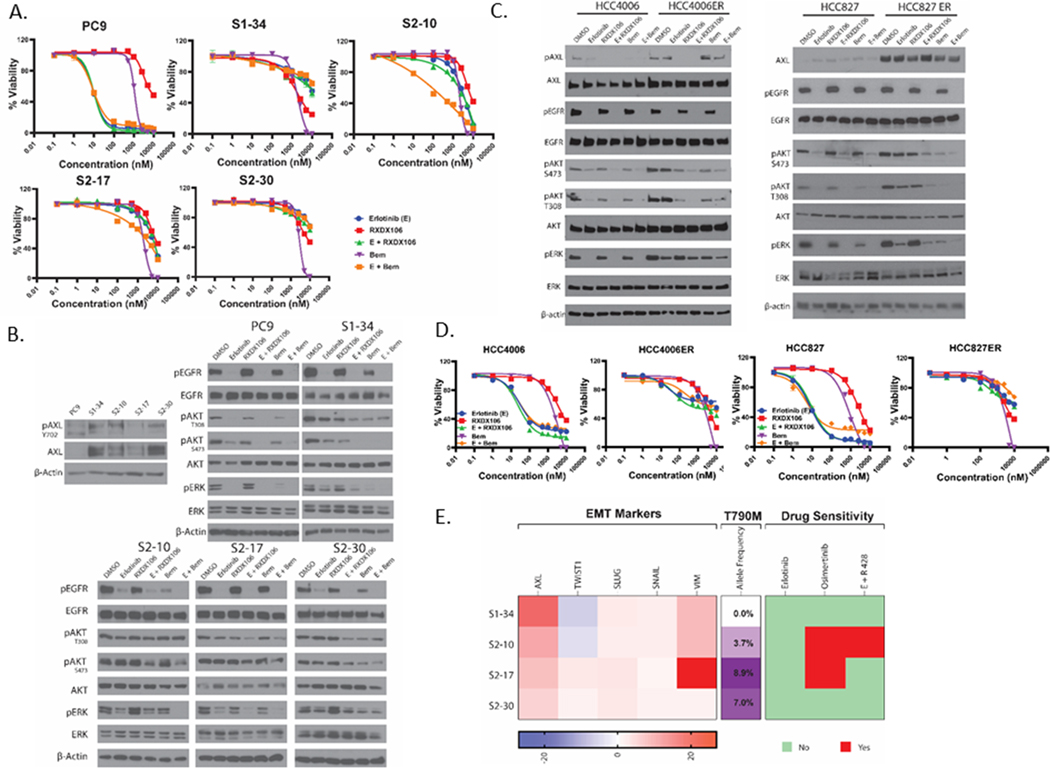
Figure 5. Effect of AXL inhibition on cell line models of erlotinib-resistance.
(A) Relative viability of PC9 and persister-derived erlotinib-resistant cells, as measured by CellTiter Glo after 72h treatment with increasing doses of the indicated drugs. For the AXL and EGFR TKI combination treatment, 1 μM of AXL inhibitor was used with increasing doses of erlotinib treatment. Data are represented as mean ± SE. (B) Immunoblot analysis of AXL expression and phosphorylation as well as alterations in downstream signaling proteins in PC9 and persister-derived erlotinib-resistant cells with or without treatment with 1μM of the indicated drugs for 1h. (C) Immunoblot assay showing changes in downstream signaling proteins in HCC4006 and HCC827 cells and their erlotinib-resistant derivative cells HCC4006ER and HCC827ER, upon treatment with 1μM of the indicated drugs for 1h. (D) Relative viability of HCC4006 and HCC827 parental and ER cells as measured by CellTiter Glo after 72h treatment with increasing doses of the indicated drugs. For the AXL and EGFR TKI combination treatment, 1 μM of AXL inhibitor was used with increasing doses of erlotinib treatment. Data are represented as mean ± SE. (E) Characterization of the persister-derived resistant clones is summarized here, highlighting differences in RNA expression of various EMT markers, allele frequencies of the T790M “gatekeeper” mutation, and sensitivity to different EGFR and AXL TKIs. All data shown are representative of three independent experiments.

