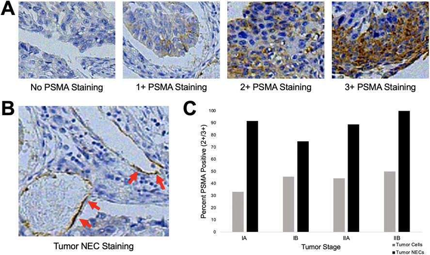
Figure 1.
Immunohistochemical analysis of PSMA expression in a cohort of patients with surgically resectable pulmonary squamous cell carcinoma. A. Representative staining intensities of pulmonary squamous cell carcinoma specimens scored as 0, no staining; 1+, weak staining; 2+, moderate staining; and 3+, strong staining. B. Representative image of PSMA-positive tumor neovascular endothelial (NEC) staining. C. Percent of specimens with PSMA expression in tumor cells or tumor NECs, stratified by final pathologic stage. PSMA was robustly expressed at all stages of disease and there were no significant differences by stage.

