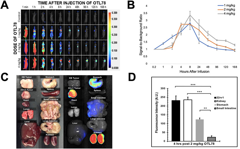
Figure 3.
Dosing, timing, and biodistribution of OTL78 in mice bearing flank xenografts. Mice bearing 22rv1 and KB flank xenografts were administered OTL78 at increasing dosing levels then imaged with the Pearl Trilogy in vivo Imaging System. A. Representative images of mice at various times after intravenous drug delivery. B. Signal-to background ratios (SBRs) were obtained for each dosing level and plotted over time from drug delivery. C. Four hours after delivery of OTL78 at 2 mg/kg, mice bearing 22rv1 and KB xenografts were euthanized to determine drug biodistribution. Fluorescence of organs and tumors were obtained using the Pearl Trilogy. D. Bar graph demonstrating fluorescence of flank tumors as compared to other organs.

