Figure 6.
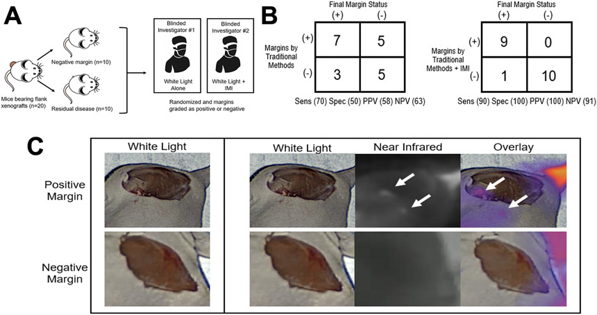
IMI with OTL78 improves identification of residual disease after resection of pulmonary squamous cell carcinoma xenografts. A. Schematic of study design. Mice bearing H1264 and H513 flank xenografts (n=10/cell line) were randomized into resection with residual disease or complete (R0) resection. All mice were administered 2 mg/kg OTL78 and mice were sacrificed 4 hours after OTL78 administration. Two blinded surgeons were then asked to grade margins as positive or negative, without knowledge of the number of positive or negative margins. One investigator could use only traditional methods (visual inspection and finger palpation) and the other could use traditional methods aided by intraoperative imaging with OTL78. B. Summary of the accuracy of margins assessed by traditional methods alone compared to those assessed with the aid of OTL78 IMI. C. Representative images of positive and negative margins as seen by the surgeon using traditional methods (left column) and the one aided by IMI with OTL78 (rightmost three columns).

