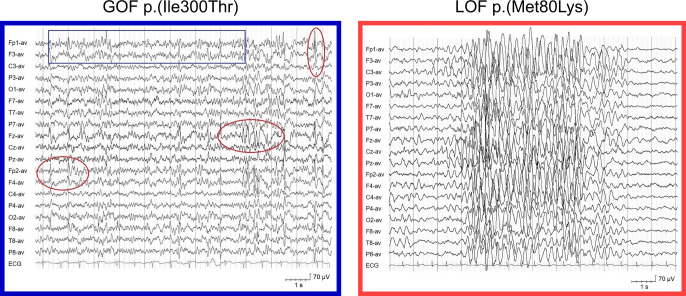
Fig. 5. Representative electroencepholagram from patients harbouring a GOF or LOF variant.
Interictal EEG showed a disorganized background activity with intermixed 12–15 Hz component (blue boxes) and multifocal epileptiform abnormalities (red circles) in patients with gain-of-function variants and normal/slightly delayed background activity, with generalized high amplitude 3–4 Hz activity/spike and slow waves (dotted lines) in patients with loss-of-function variants.

