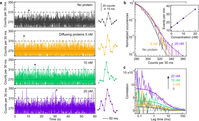
Fig. 3. Label-free detection of single diffusing proteins across the UV horn antenna.
55% sucrose was added to the buffer to slow down the protein diffusion by 30× and ease observing the autofluorescence bursts. a Autofluorescence time traces with increasing β-galactosidase-streptavidin concentrations. The binning time is 30 ms. The stars indicate selected autofluorescence bursts which are displayed on the right panel with 15 ms bin time to better view individual bursts. b Normalized photon counts histograms computed over the full trace duration of 200 s. The thick lines are numerical fits using the sum of a Gaussian and an exponential distribution to account for the noise and the autofluorescence bursts respectively. The inset shows the number of detected events per minute above the threshold corresponding to 2.5× the standard deviation of the noise when no protein is present (dashed lines at 310 counts in (a). c FCS correlation functions of the traces displayed in (a). The thick lines are numerical fits.

